Welcome
By selecting the Yes, leave Opcaseportalmy.com option you understand that you are leaving the website and that you will be redirected to another Internet site belonging to third parties, for whose content or opinions Amgen Malaysia is not responsible and makes no representations regarding such content or its accuracy. By selecting this option and accessing linked third-party sites, you understand that you do so at your own risk.
The website to which you will be redirected may be designed exclusively for residents of a particular country or countries, as advertised on that website. As a consequence, the website to which you will be directed may contain information on pharmaceutical products or indications that are not approved in Malaysia. If you reside in a country other than the one to which such website is directed, please contact your local Amgen affiliate for the correct information on products in your country of residence.


Case contributed by Prof. Datuk Dr. Sabarul Afian Mokhtar, Senior Consultant Orthopaedic Surgeon (Spine Surgery)
Hospital Canselor Tuanku Muhriz Universiti Kebangsaan Malaysia
Patient information
Age: 62 years
Sex: Female
-
History
- Menopause at 52 years old
- No history of cardiovascular events
- No family history of osteoporosis
Presenting complaint
- Patient was referred for lower back pain
- She was a passenger at the back seat of a car, who sustained the injury when the car hit a speed bump
- Diagnosed with vertebral fracture at L1 upon admission
-
Physical Examination
Weight: 58 kg
Height: 154 cm
BMI: 24.5 kg/m2
Specific
- No features of neurological deficit
- No features of Cushing syndrome
-
Investigation
Blood investigations
- All blood investigations were normal
-
Treatment
- Treatment-naïve
Pharmacological
- Romosozumab therapy for 1 year, followed by transition to Denosumab 6-monthly
Non-pharmacological
- Apart from osteoporosis treatment, the patient was on a Thoracic-Lumbar-Sacral Orthosis (TLSO) brace to immobilize the fracture for 3 months until pain relief
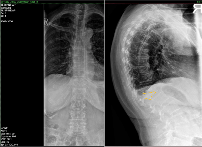
Spine X-ray upon admission
Spine X-ray showing wedge compression fracture of vertebrae
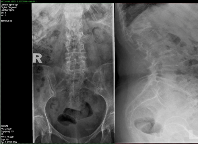
Lumbosacral X-ray after 12 months
No adjacent new fractures observed after 12 months with romosozumab therapy
-
Follow Up
Upon completion of Romosozumab therapy for 1 year, patient was transitioned to Denosumab
6-monthly -
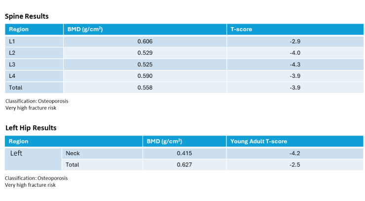
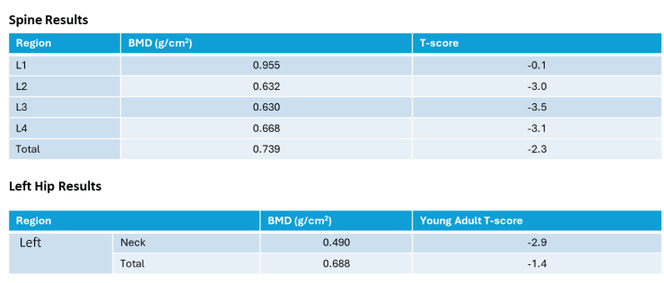
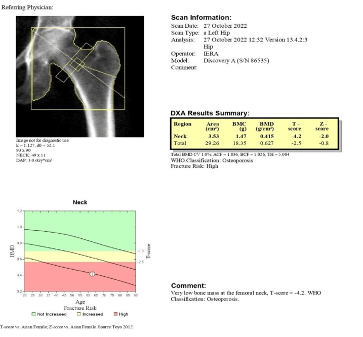
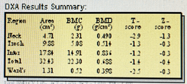
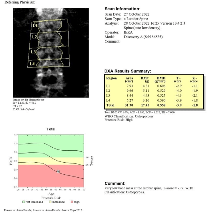
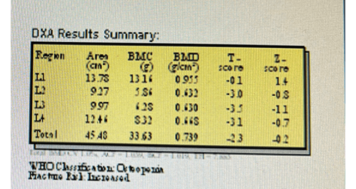
Results- DXA Scan
Upon admission
After 1 year of Romosozumab therapy
Left Hip Upon Admission
Left Hip Upon Completing 12 Months of Romosozumab
Spine Upon Admission
Spine Upon Completing 12 Months of Romosozumab
-
Summary
- Patient sustained vertebral fracture at L1
- 1st DXA scan done upon admission
- Started on monthly subcutaneous Romosozumab after fracture
2nd DXA scan done after 12 months of Romosozumab therapy
- Spine BMD changes: [(0.739-0.558)/0.558] X 100% = 32.4%
- Hip BMD changes: [(0.688 - 0.627)/ 0.627] X 100 = 9.7%
- After 1 year of Romosozumab therapy, patient was switched to subcutaneous Denosumab
6-monthly therapy

-
Estimated Fracture Risk Reduction Associated With BMD Improvement1

-
References
1. Bouxsein ML, Eastell R, Lui LY, et al.; FNIH Bone Quality Project. Change in Bone Density and Reduction in Fracture Risk: A Meta-Regression of Published Trials. J Bone Miner Res. 2019 Apr;34(4):632-642.
Abbreviations
BMD, bone mineral density; DXA, dual-energy X-ray absorptiometry; L1, first lumbar vertebrae; L2, second lumbar vertebrae; L3, third lumbar vertebrae; L4, fourth lumbar vertebrae
-
Important Safety Information:Osteoporosis Case BinderThis curated collection of patient case studies aims to provide healthcare practitioners in the area of bone health, with a comprehensive understanding of risk-based treatment approach in the long-term management of patients living with osteoporosis.The patient cases were contributed by bone health experts from respective clinical setting, incorporating evidence-based discussions, guidelines and clinical considerations in the individualised treatment sequencing plans of different real-world scenarios..Please review full product information before prescribingFor Healthcare Professionals Only.Please refer to full prescribing information prior to administrationFor information on Amgen products or to report an adverse event involving an Amgen product, please contact Medical Information at 1800 818 227 or medinfo.JAPAC@amgen.comSC-MAL-CP-00128-0425Endorsed By: